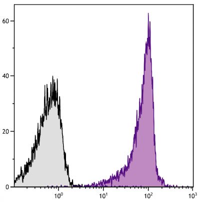
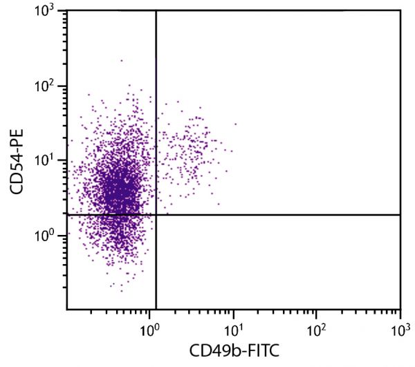
Rat Anti-Mouse CD54-PE (YN1/1.7.4)
Cat. No.:
1701-09
PE Anti-Mouse CD54 antibody for use in flow cytometry assays.
$236.00

| Clone | YN1/1.7.4 |
|---|---|
| Isotype | Rat (Fischer 344) IgG2bκ |
| Isotype Control | Rat IgG2b-PE (KLH/G2b-1-2) |
| Specificity | Mouse/Porcine CD54 |
| Alternative Names | ICAM-1, Ly-47 |
| Description | CD54, a member of the immunoglobulin superfamily of cell surface receptors and also known as Intercellular Adhesion Molecule-1 (ICAM-1), is a type I transmembrane glycoprotein that mediates important cell/cell interactions. CD54 is expressed on endothelial cells, dendritic cells, keratinocytes, and lymphocytes. Its expression is upregulated by inflammatory mediators such as IFN γ, IL-1, TNF-α, lipopolysaccharide, and phorbol esters. Endothelial CD54 contributes to the extravasation of leukocytes from blood vessels, particularly in areas of inflammation. CD54 on antigen-presenting cells contributes to antigen-specific T-cell activation, presumably by enhancing interactions between T cell and antigen-presenting cells. These adhesion reactions are mediated via the binding of CD54 to its major ligands, LFA-1 and Mac-1. |
| Immunogen | Mouse NS-1 cells |
| Conjugate | PE (R-phycoerythrin) |
| Buffer Formulation | Phosphate buffered saline containing < 0.1% sodium azide and a stabilizer |
| Clonality | Monoclonal |
| Concentration | 0.1 mg/mL |
| Volume | 1.0 mL |
| Recommended Storage | 2-8°C; Avoid exposure to light; Do not freeze |
| Applications |
Flow Cytometry – Quality tested 9-11 Immunohistochemistry-Frozen Sections – Reported in literature 3,12-14 Immunocytochemistry – Reported in literature 4 Western Blot – Reported in literature 6-8 Immunoprecipitation – Reported in literature 1 Blocking – Reported in literature 1,2 Purification – Reported in literature 5 |
| RRID Number | AB_2795154 |
| Gene ID |
15894 (Mouse) 396750 (Porcine) |
| Gene ID Symbol |
Icam1 (Mouse) ICAM1 (Porcine) |
| Gene ID Aliases | CD54; Icam-1; Ly-47; MALA-2 |
| UniProt ID |
P13597 (Mouse) |
| UniProt Name |
ICAM1_MOUSE (Mouse) |
Documentation
Certificate of Analysis Lookup
Enter the Catalog Number and Lot Number for the Certificate of Analysis you wish to view
- 1. Takei F. Inhibition of mixed lymphocyte response by a rat monoclonal antibody to a novel murine lymphocyte activation antigen (MALA-2). J Immunol. 1985;134:1403-7. (Immunogen, FC, IP, Block)
- 2. Kumasaka T, Quinlan WM, Doyle NA, Condon TP, Sligh J, Takei F, et al. Role of the intercellular adhesion molecule-1(ICAM-1) in endotoxin-induced pneumonia evaluated using ICAM-1 antisense oligonucleotides, anti-ICAM-1 monoclonal antibodies, and ICAM-1 mutant mice. J Clin Invest. 1996;97:2362-9. (Block)
- 3. Song J, Lokmic Z, Lämmermann T, Rolf J, Wu C, Zhang X, et al. Extracellular matrix of secondary lymphoid organs impacts on B-cell fate and survival. Proc Natl Acad Sci USA. 2013;110(31):E2915-24. (IHC-FS)
- 4. Bosch B, Heipertz EL, Drake JR, Roche PA. MHC class II-peptide complexes arrive at the plasma membrane in cholesterol-rich microclusters. J Biol Chem. 2013;288:13236-42. (ICC)
- 5. Krauss K, Altevogt P. Integrin leukocyte function-associated antigen-1-mediated cell binding can be activated by clustering of membrane rafts. J Biol Chem. 1999;274:36921-7. (Purification)
- 6. Muntasell A, Berger AC, Roche PA. T cell-induced secretion of MHC class II–peptide complexes on B cell exosomes. EMBO J. 2007;26:4263-72. (WB)
- 7. Ramachandra L, Qu Y, Wang Y, Lewis CJ, Cobb BA, Takatsu K, et al. Mycobacterium tuberculosis synergizes with ATP to induce release of microvesicles and exosomes containing major histocompatibility complex class II molecules capable of antigen presentation. Infect Immun. 2010;78:5116-25. (WB)
- 8. Chen L, Lin S, Amin S, Overbergh L, Maggiolino G, Chan LS. VCAM-1 blockade delays disease onset, reduces disease severity and inflammatory cells in an atopic dermatitis model. Immunol Cell Biol. 2010;88:334-42. (WB)
- 9. Zaru R, Mollahan P, Watts C. 3-Phosphoinositide-dependent kinase 1 deficiency perturbs toll-like receptor signaling events and actin cytoskeleton dynamics in dendritic cells. J Biol Chem. 2008;283:929-39. (FC)
- 10. Podgrabinska S, Kamalu O, Mayer L, Shimaoka M, Snoeck H, Randolph GJ, et al. Inflamed lymphatic endothelium suppresses dendritic cell maturation and function via Mac-1/ICAM-1-dependent mechanism. J Immunol. 2009;183:1767-79. (FC)
- 11. Jacobsen J, Haabeth OW, Tveita AA, Schjetne KW, Munthe LA, Bogen B. Naive idiotope-specific B and T cells collaborate efficiently in the absence of dendritic cells. J Immunol. 2014;192:4174-83. (FC)
- 12. Hoehn D, Sun L, Sucosky P. Role of pathologic shear stress alterations in aortic valve endothelial activation. Cardiovasc Eng Technol. 2010;1:165-78. (IHC-FS, Porcine Reactivity)
- 13. Sun L, Chandra S, Sucosky P. Ex vivo evidence for the contribution of hemodynamic shear stress abnormalities to the early pathogenesis of calcific bicuspid aortic valve disease. PLoS One. 2012;7(10):e48843. (IHC-FS, Porcine Reactivity)
- 14. Sun L, Rajamannan NM, Sucosky P. Defining the role of fluid shear stress in the expression of early signaling markers for calcific aortic valve disease. PLoS One. 2013;8(12):e84433. (IHC-FS, Porcine Reactivity)
See All References
Related Products
Check items to add to the cart or