Mouse Anti-Rabbit RLA-DR-BIOT (RDR34)
Cat. No.:
4070-08
Biotin Anti-Rabbit RLA-DR antibody for use in flow cytometry, immunohistochemistry, and immunoprecipitation assays.
$347.00
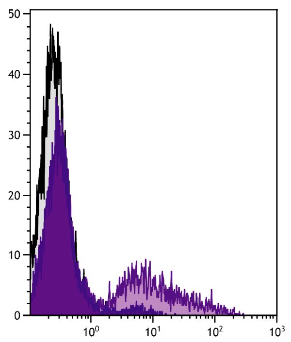

| Clone | RDR34 |
|---|---|
| Isotype | Mouse (BALB/c) IgG2bκ |
| Isotype Control | Mouse IgG2b-BIOT (A-1) |
| Specificity | Rabbit RLA-DR |
| Alternative Names | MHC Class II, leukocyte antigen |
| Description | The monoclonal antibody RDR34 reacts with RLA-DR-transfected cells but not with RLA-DQ-transfected cells. In normal adult rabbits, the antibody reacts with 50-60% of peripheral blood leukocytes and splenocytes. It has also been reported that the antibody stains lymphocytes in the thymic medulla and some cells in the cortex, periportal cells in the liver, and synovial lining cells (e.g., infiltrating macrophages) in inflamed synovium. DRα transcripts have been detected in appendix, bone marrow, spleen, and to a lesser extent in lymph nodes, lung, liver, and other organs. |
| Immunogen | Rabbit leukemic B cell line |
| Conjugate | BIOT (Biotin) |
| Buffer Formulation | Phosphate buffered saline containing < 0.1% sodium azide |
| Clonality | Monoclonal |
| Concentration | 0.5 mg/mL |
| Volume | 1.0 mL |
| Recommended Storage | 2-8°C |
| Applications |
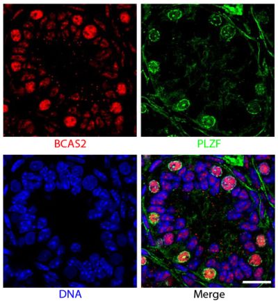
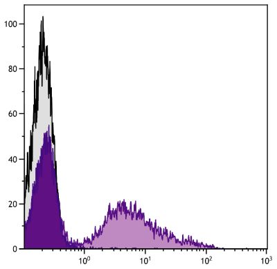
Flow Cytometry – Quality tested 1,2 Immunohistochemistry-Frozen Sections – Reported in literature 3-5 Immunohistochemistry-Whole Mount – Reported in literature 6 Immunoprecipitation – Reported in literature 1 |
| RRID Number | AB_2795993 |
| Gene ID |
100328929 (Rabbit) |
| Gene ID Symbol |
RLA-DR-ALPHA (Rabbit) |
Documentation
Certificate of Analysis Lookup
Enter the Catalog Number and Lot Number for the Certificate of Analysis you wish to view
- 1. Spieker-Polet H, Sittisombut N, Yam P, Knight KL. Rabbit major histocompatibility complex. IV. Expression of major histocompatibility complex class II genes. J Immunogenet. 1990;17:123-32. (Immunogen, FC, IP)
- 2. Sawasdikosol S, Hague BF, Zhao TM, Bowers FS, Simpson RM, Robinson M, et al. Selection of rabbit CD4-CD8- T cell receptor-γ/δ cells by in vitro transformation with human T lymphotropic virus-I. J Exp Med. 1993;178:1337-45. (FC)
- 3. Wilkinson JM, McDonald G, Smith S, Galea-Lauri J, Lewthwaite J, Henderson B, et al. Immunohistochemical identification of leucocyte populations in normal tissue and inflamed synovium of the rabbit. J Pathol. 1993;170:315-20. (IHC-FS)
- 4. Vinuesa M, Bassan N, Roma S, Pérez F. Immunopathological modifications in the rectal mucosa from an animal model of food allergy. Rev Esp Enferm Dig. 2005;97:629-36. (IHC-FS)
- 5. Brignole-Baudouin F, Desbenoit N, Hamm G, Liang H, Both J, Brunelle A, et al. A new safety concern for glaucoma treatment demonstrated by mass spectrometry imaging of benzalkonium chloride distribution in the eye, an experimental study in rabbits. PLoS One. 2012;7(11):e50180. (IHC-FS)
- 6. Huang W, Chamberlain CG, Sarafian RY, Chan-Ling T. MHC class II expression by β2 integrin (CD18)-positive microglia, macrophages and macrophage-like cells in rabbit retina. Neuron Glia Biol. 2008;4:285-94. (IHC-WM)
See More


