Mouse Anti-Human CD43-BIOT (DF-T1)
Cat. No.:
9620-08
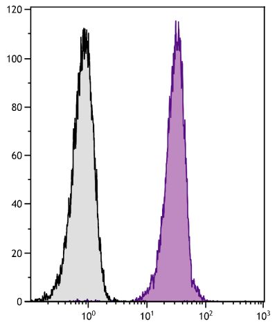
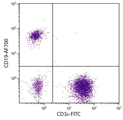
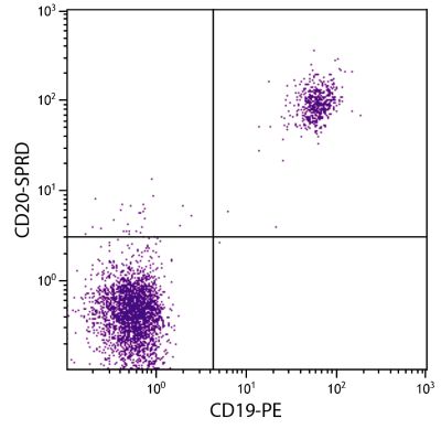
Biotin Anti-Human CD43 antibody for use in flow cytometry, immunohistochemistry / immunocytochemistry, western blot, and immunoprecipitation assays.
$185.00
| Clone | DF-T1 |
|---|---|
| Isotype | Mouse IgG1κ |
| Isotype Control | Mouse IgG1-BIOT (15H6) |
| Specificity | Human CD43 |
| Alternative Names | gpL115, leukocyte sialoglycoprotein |
| Description | CD43, a member of the sialomucin family of cell surface receptors, is a 95-135 kDa type I transmembrane glycoprotein whose variability in molecular weight is dependent upon the nature of the extracellular O-glycans. It is the major sialoglycoprotein on thymocytes, T cells, and neutrophils. It is also present on activated B cells, plasma cells, NK cells, granulocytes, monocytes, macrophages, platelets, and bone marrow hematopoietic stem cells. CD43, which binds to ICAM-1 (CD54), appears to function as an anti-adhesion molecule, inhibiting T cell interactions including T cell killing, and by increasing the threshold for T cell activation. |
| Immunogen | KG-1 cells |
| Conjugate | BIOT (Biotin) |
| Buffer Formulation | Phosphate buffered saline containing < 0.1% sodium azide |
| Clonality | Monoclonal |
| Concentration | Lot specific |
| Volume | 1.0 mL |
| Recommended Storage | 2-8°C |
| Applications |
Flow Cytometry – Quality tested 1,5,6 Immunohistochemistry-Frozen Sections – Reported in literature 1 Immunohistochemistry-Paraffin Sections – Reported in literature 1,2 Immunocytochemistry – Reported in literature 1 Immunoprecipitation – Reported in literature 3,4 Western Blot – Reported in literature 1,3,5,6 Adhesion – Reported in literature 3,4 |
| RRID Number | AB_2797009 |
| Gene ID |
6693 (Human) |
| Gene ID Symbol |
SPN (Human) |
| Gene ID Aliases | CD43; GALGP; GPL115; LSN |
| UniProt ID |
P16150 (Human) |
| UniProt Name |
LEUK_HUMAN (Human) |
Documentation
Certificate of Analysis Lookup
Enter the Catalog Number and Lot Number for the Certificate of Analysis you wish to view
- 1. Stross WP, Warnke RA, Flavell DJ, Flavell SU, Simmons D, Gatter KC, et al. Molecule detected in formalin fixed tissue by antibodies MT1, DF-T1, and L60 (Leu-22) corresponds to CD43 antigen. J Clin Pathol. 1989;42:953-61. (IHC-PS, IHC-FS, ICC, WB, FC)
- 2. Lopez-Beltran A, Santamaria M, Garcia-Cozar FJ, Millan JM, Pena J, Molina IJ. CD43 is detected in paraffin-embedded sections of non-haemopoietic malignant tumours. In: Schlossman SF, Boumsell L, Gilks W, Harlan JM, Kishimoto T, Morimoto C, et al, editors. Leukocyte Typing V: White Cell Differentiation Antigens. Oxford: Oxford University Press; 1995. p. 1712-13. (IHC-PS)
- 3. Remold-O'Donnell E. CD43 cluster report. In: Schlossman SF, Boumsell L, Gilks W, Harlan JM, Kishimoto T, Morimoto C, et al, editors. Leukocyte Typing V: White Cell Differentiation Antigens. Oxford: Oxford University Press; 1995. p. 1697-1701. (IP, WB, Adhesion)
- 4. Axelsson B, Etemad R, Nordlund R. Biochemical and funtional characterization of the workshop adhesion structure subpanel 9 (CD43) mAb. In: Schlossman SF, Boumsell L, Gilks W, Harlan JM, Kishimoto T, Morimoto C, et al, editors. Leukocyte Typing V: White Cell Differentiation Antigens. Oxford: Oxford University Press; 1995. p. 1708-9. (IP, Adhesion)
- 5. Parent D, Remold-O'Donnell E. Reactivity of mAb with CD43 isoforms. In: Schlossman SF, Boumsell L, Gilks W, Harlan JM, Kishimoto T, Morimoto C, et al, editors. Leukocyte Typing V: White Cell Differentiation Antigens. Oxford: Oxford University Press; 1995. p. 1701-2. (WB, FC)
- 6. Chorvath B, Duraj J, Mellors A, Hunakova L, Sedlak J. Cell surface leukosialin (CD43) as a substrate for Parteurella haemolytica O-glycoprotease. In: Schlossman SF, Boumsell L, Gilks W, Harlan JM, Kishimoto T, Morimoto C, et al, editors. Leukocyte Typing V: White Cell Differentiation Antigens. Oxford: Oxford University Press; 1995. p. 1705-6. (WB, FC)
See More


