
Share a link
Western Blotting Troubleshooting
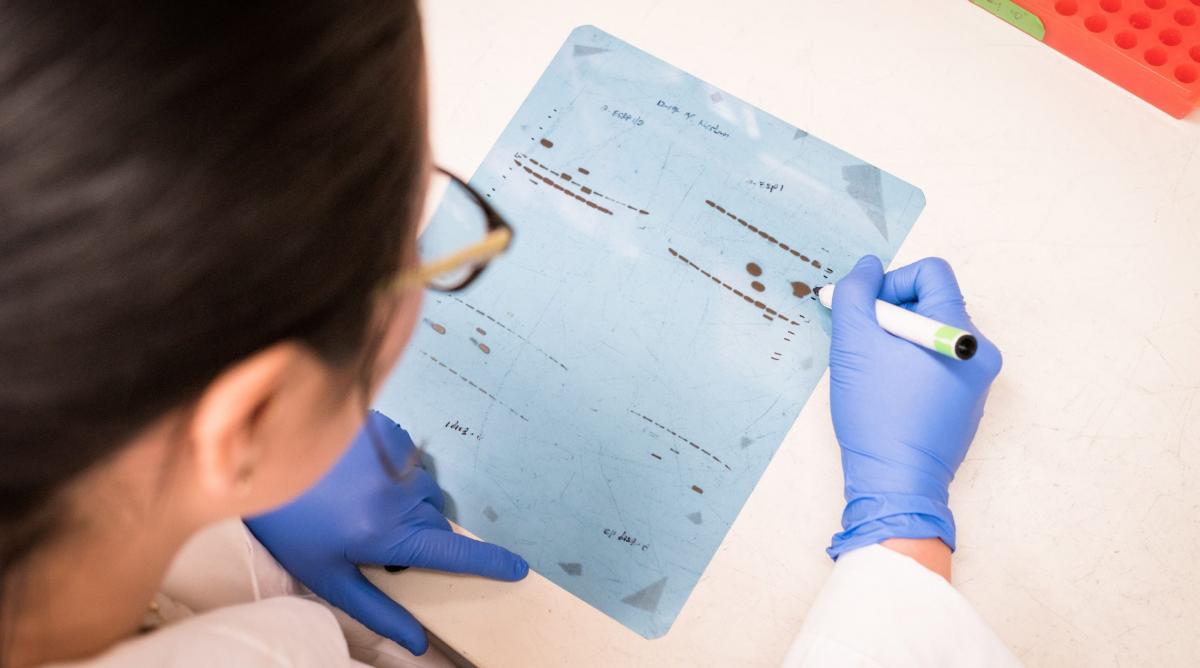
Western blotting provides researchers with an image of one or more proteins within a heterogeneous sample. Yet, despite being used extensively, Western blotting may not always go to plan. Here, we've listed some common Western blotting issues and suggested possible solutions.

I can't see any bands / the bands are very faint
Poor Western blot signal is often due to samples being mishandled or antibodies not being properly optimized for target detection.
| Cause | Solution |
|---|---|
|
The sample has degraded |
Keep samples on ice; add protease and/or phosphatase inhibitors to the sample lysis buffer; avoid freeze-thaw cycles |
|
The target of interest is only expressed at low levels in the chosen sample type |
Refer to sites such as UniProt, PAXdb, or proteinatlas.org, and to antibody manufacturers’ datasheets, for information about target expression; try loading more sample per lane; consider enriching the target via immunoprecipitation |
|
The primary antibody has poor affinity for the target |
Check that the primary antibody is validated for Western blot, as well as for the chosen species; establish whether the primary antibody recognizes native or denatured protein; run a positive control to check antibody performance |
|
The primary and secondary antibodies were incompatible |
Ensure that an appropriate secondary antibody was used for detection (e.g., if the primary antibody was raised in mouse, an anti-mouse secondary antibody should be used) |
|
The antibodies were too dilute |
Titrate primary and secondary antibody reagents to optimize concentrations |
We offer a broad range of antibodies that have been validated for Western blotting, which includes directly-labeled primary antibody reagents.
There are too many bands / the bands are the wrong size
The presence of unexpected bands on a Western blot can be indicative of sample degradation, but it can also represent a genuine result.
| Cause | Solution |
|---|---|
|
The sample has degraded |
Keep samples on ice; add protease and/or phosphatase inhibitors to the sample lysis buffer; avoid freeze-thaw cycles |
|
The target protein exists as multiple isoforms |
Refer to sites such as UniProt, PAXdb, or proteinatlas.org, and to antibody manufacturers’ datasheets, for information about target expression; consider using isoform-specific primary antibodies, if available |
|
Post-translational modifications (PTMs) have altered the predicted molecular weight of the target |
Determine whether the target undergoes PTMs such as phosphorylation, glycosylation, or cleavage |
|
Primary antibodies are binding non-specifically to other proteins |
Switch to using different primary antibodies; use a blocking peptide to differentiate between specific and non-specific bands; re-optimize the blocking conditions |
|
Fluorophore-labeled secondary antibodies are cross-reacting in a multiplexed Western blot |
Test each secondary antibody separately to verify performance; use secondary antibodies from the same host species; switch to using cross-adsorbed secondary antibodies |
Many of our secondary antibodies are cross-adsorbed against multiple species, including products for detecting mouse, rat, human, goat, and rabbit IgG.
My Western blot has high background
High background across a Western blot is frequently a product of poor blocking, inadequate washing, or, in the case of chemiluminescent detection, over-exposure.
| Cause | Solution |
|---|---|
|
The blot was not blocked properly |
Optimize the blocking conditions; switch to using a different blocking buffer; include the blocking agent in antibody diluent solutions |
|
Washing was insufficient |
Increase the number, duration, and volume of wash steps; consider adding a low concentration of detergent (e.g., 0.1% Tween® 20) to wash buffers |
|
The antibody concentrations were too high |
Titrate primary and secondary antibody reagents to optimize concentrations |
|
Secondary antibodies have bound non-specifically to the membrane |
Avoid using milk for blocking, especially when working with anti-goat secondary antibodies; run a secondary antibody only control |
|
The blot was over-exposed (chemiluminescent detection) |
Reduce the exposure time; consider using an imaging system that offers an auto-exposure function |
My Western blot looks patchy or speckled
Unexpected background features can be due to the presence of air bubbles, contamination, or blocking buffer precipitates, as well as poor handling of the membrane.
| Cause | Solution |
|---|---|
|
Air was trapped between the gel and the membrane |
Use a roller to remove air bubbles when setting up the transfer |
|
Contamination was present |
Always wash and dry blotting apparatus after use; routinely soak transfer pads and incubation trays in 100% methanol; prepare fresh buffers for every assay or store buffers short-term at 4oC if appropriate |
|
The blocking buffer contained precipitates |
Filter the blocking buffer prior to use; switch to using a different blocking buffer |
|
The membrane was compromised by handling |
Always wear gloves when handling the membrane; use blunt-edged forceps to avoid causing scratches |
|
Several membranes were incubated together in the same tray |
Always use a separate tray for each membrane |
The lanes on my Western blot are smeared
Smeared lanes are almost always due to poor sample preparation, which can lead to degradation, protein aggregation, and over-loading.
| Cause | Solution |
|---|---|
|
The sample has degraded |
Keep samples on ice; add protease and/or phosphatase inhibitors to the sample lysis buffer; avoid freeze-thaw cycles |
|
DNA has caused proteins to aggregate |
Consider adding DNase to the lysis buffer |
|
The sample was overloaded |
Check the protein concentration; reduce the amount of sample loaded per lane |
|
The sample was not solubilized properly |
Switch to using a different lysis buffer; increase the time spent sonicating or heating samples; centrifuge samples before loading them onto the gel |
|
The transfer was not set up correctly |
Use thicker filter paper and/or increase the number of sponges in the sandwich to ensure proper contact between the gel and the membrane |
While this list of troubleshooting tips is by no means complete, it highlights the critical importance of understanding the target, preparing samples carefully, and optimizing Western blotting conditions. If you need further guidance, our highly experienced technical support team is available to help.


