Share a link
Assays to Detect Apoptosis
Detecting apoptosis requires multiple approaches across early, mid, and late stages. Explore key assays, including Annexin V, mitochondrial membrane potential, caspases, and TUNEL, to better understand cell death pathways, disease mechanisms, and therapeutic applications in research.

What is apoptosis?
Apoptosis, also known as programmed cell death, is a highly controlled process for removing damaged or redundant cells from multicellular organisms. Its dysregulation has been linked to developmental abnormalities and conditions including autoimmune disorders, neurodegenerative diseases, and cancer. For this reason, many different assays have been developed to monitor apoptosis. These include methods for evaluating plasma membrane integrity, assessing mitochondrial function, and measuring caspase activity, as well as assays for identifying DNA fragmentation.
We offer various reagents that support apoptosis research, including fluorescently-labeled annexin V conjugates and primary antibodies to targets along the main apoptosis signaling pathways. In addition, we provide secondary antibodies specific to numerous species, which are available in a broad range of conjugate options to maximize flexibility for experimental design.
Understanding Apoptosis
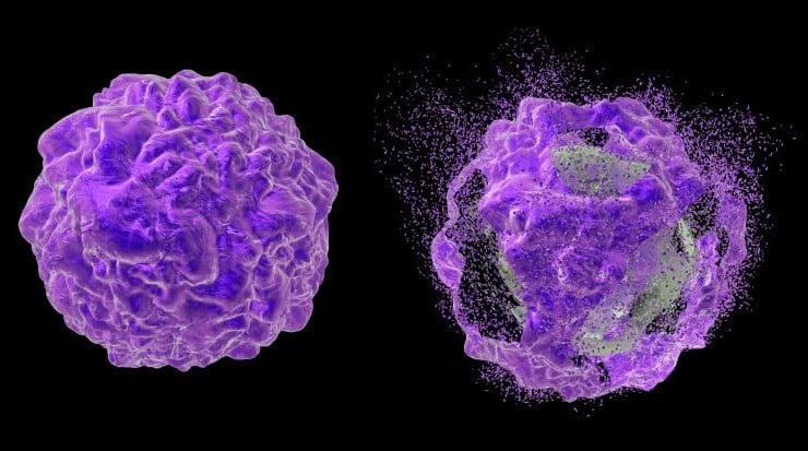
Apoptosis is responsible for cell loss during embryogenesis, development of the nervous system, and normal cell turnover, as well as for establishing a functional immune system through negative selection. It is distinct from necrosis, or accidental cell death, in that it is characterized by specific morphological changes. These include nuclear condensation, cell shrinkage, and membrane blebbing, as well as DNA fragmentation and cleavage or degradation of various cellular proteins.
- Apoptosis induction pathways
Apoptosis is induced through three main pathways: the extrinsic pathway, the intrinsic pathway, and the perforin/granzyme pathway. The extrinsic pathway, which begins outside of the cell, is initiated by ligand binding to cell surface receptors, commonly known as death receptors, leading to activation of the caspase cascade. Well-known cell death receptors include the Fas receptor (FasR) and tumor necrosis factor receptor 1 (TNFR1), which are triggered by the Fas ligand (FasL) and tumor necrosis factor α (TNF-α), respectively.
The intrinsic pathway is induced by developmental cues and cellular stresses, such as DNA damage, accumulation of misfolded proteins, oxidative stress, hypoxia, and nutrient deprivation. It leads to permeabilization of mitochondrial outer membranes, resulting in diminished ATP synthesis, perturbation to the mitochondrial respiratory chain, and the release of cytotoxic proteins such as cytochrome C, apoptosis inducing factor (AIF), and endonuclease G (EndoG).
The perforin/granzyme pathway is crucial for cytotoxic T cells to eliminate aberrant cells, such as tumor cells and cells infected with viruses or intracellular parasites. When a cytotoxic T cell encounters a target cell, the resultant signaling cascade causes the release of granules containing perforin, a membrane pore-forming protein, and granzymes such as Granzyme A and Granzyme B, which enter the target cell to induce apoptosis. Notably, different granzymes trigger apoptosis via different mechanisms.
- Stages of apoptosis
Apoptosis is a multi-step process that can be broadly divided into early, mid, and late-stage cellular events. Early apoptosis is characterized by the translocation of phosphatidylserine (PS), a key component of the phospholipid bilayer, to the outer leaf of the plasma membrane, where it serves to trigger phagocytosis. Other early apoptosis events include the loss of mitochondrial membrane potential, the mitochondrial release of cytochrome C and ATP into the cytosol, and the activation of initiator caspases, such as caspase 8 and caspase 9.
Mid-stage apoptosis involves activation of executioner caspases, such as caspases 3, 6, and 7, which are responsible for cleaving various cellular proteins. Their targets include the nuclear enzyme poly (ADP-ribose) polymerase (PARP), an essential DNA repair protein, and lamin A/C, a key structural component of the nuclear membrane. Other mid-stage apoptosis events include cell shrinkage, cytoskeleton collapse, and the activation of nucleases.
During late-stage apoptosis, DNA fragmentation is accompanied by nuclear collapse and the formation of apoptotic bodies, which are engulfed by phagocytes. Assays for monitoring apoptosis target many different events, making it important to select the right approach for your specific application.
What are the key cell apoptosis assays?
Because apoptosis is a highly complex process, researchers must often perform multiple assays to determine its occurrence. The following are some of the most widely used methods for studying apoptosis -
- Annexin V assays
Annexin V is a Ca2+-dependent phospholipid-binding protein with a high affinity for phosphatidylserine (PS). In normal, healthy cells, PS is found on the cytoplasmic side of the plasma membrane. However, during early apoptosis, PS translocates to the cellular surface, where it can be detected using fluorescently-labeled Annexin V conjugates for techniques such as flow cytometry and immunocytochemistry (ICC). To distinguish apoptotic cells from necrotic cells, Annexin V is typically used in tandem with reagents such as propidium iodide (PI) and 7-aminoactinomycin (7-AAD), which can only enter cells with leaky membranes.
- Mitochondrial membrane potential assays
Mitochondria are the main cellular powerhouses, essential for ATP synthesis, calcium homeostasis, and steroid metabolism. During early apoptosis, sustained opening of mitochondrial permeability transition pores (MPTPs) leads to a loss of mitochondrial membrane potential (MMP) that can be measured using cell permeable dyes such as tetramethylrhodamine ethyl ester (TMRE). In normal cells, TMRE accumulates in mitochondria, where it produces orange-red fluorescence when excited at 550 nm. In apoptotic cells, MMP collapse prevents the accumulation of TMRE, which corresponds with a lack of fluorescent signal.
- Cytochrome C release assays
The loss of mitochondrial membrane potential during early apoptosis results in the release of cytochrome C into the cytosol. Here, cytochrome C interacts with apoptotic protease activating factor-1 (Apaf-1) to form a complex known as the apoptosome, which activates caspase-9 and triggers a cascade of events that lead to the cell being dismantled. Cytochrome C can be detected using highly specific primary antibodies in techniques including immunocytochemistry (ICC) and Western blot analysis of cytosolic and mitochondrial cell extracts.
- Caspase assays
Caspases are a family of protease enzymes that serve as critical mediators of apoptosis. They are divided into two groups: the initiator caspases, such as caspases 2, 8, 9, 10, and 12, and the executioner caspases, such as caspases 3, 6, and 7. Initiator caspases activate executioner caspases, enabling them to cleave various cellular proteins to facilitate apoptosis. Caspase assays measure caspase activity within cells by detecting specific cleavage products with techniques including Western blot, ELISA, and immunohistochemistry (IHC).
- TUNEL assays
TUNEL (terminal deoxynucleotidyl transferase dUTP nick end labeling) assays are used for monitoring DNA fragmentation in late-stage apoptosis. They work by attaching labeled nucleotides (e.g., BrdUTP, biotin-dUTP, or fluorescein-dUTP) to the 3' ends of damaged DNA, where they are detected using specific antibodies. The more nucleotides that are incorporated into the DNA, the higher the signal, meaning the further apoptosis has progressed. TUNEL assays are commonly performed using techniques such as immunocytochemistry (ICC), immunohistochemistry (IHC), and flow cytometry.
What techniques are used for apoptosis detection?
Many different detection techniques are used for studying apoptosis, including those described in the Key Cell Apoptosis Assays section above. Deciding which technique to use will depend on your sample type and target of interest. For example, if your intention is to detect phosphatidylserine (PS) at the cell surface, you might consider flow cytometry for a cell suspension, immunocytochemistry (ICC) for adherent cells, or immunohistochemistry (IHC) for an excised tissue section. In contrast, if you are handling cell lysates, techniques such as Western blotting and ELISA are more suitable. For lysates in which the target of interest has low abundance, immunoprecipitation (IP) can be used for enrichment.
What are some considerations for apoptosis assay selection?
When selecting an apoptosis assay, one of the first factors to consider is whether you need to focus on a particular apoptosis induction pathway. You will also want to think about which stage of apoptosis you are most interested in, since early-, mid-, and late-stage apoptosis are each characterized by different events. Other considerations for assay selection include which method to use and whether you should run multiple methods in parallel to increase confidence in your results. For example, measuring both TMRE staining and cytochrome C release can help to confirm mitochondrial dysfunction. Lastly, it is essential that you identify high-quality reagents to support your studies and validate them for use in your specific model system before embarking on data-driven research.
Applications in Research and Medicine
Dysregulated apoptosis is known to contribute to developmental abnormalities such as malformations of digits and cardiac valves, as well as to neurodegenerative diseases including Parkinson's disease, Alzheimer's disease, Huntington's disease, and amyotrophic lateral sclerosis (ALS). Apoptosis is also implicated in autoimmune disorders, cancer, stroke, and chronic viral infections, making it a main focus for many different fields of research. Well-validated antibodies, kits, and other reagents are essential for studying the role of apoptosis in health and disease. SouthernBiotech offers various products to support apoptosis research, including fluorescently-labeled annexin V conjugates and primary antibodies to key apoptosis signaling molecules. We also provide secondary antibodies that can be used for signal amplification.


