
Share a link
Introduction To Immunohistochemistry
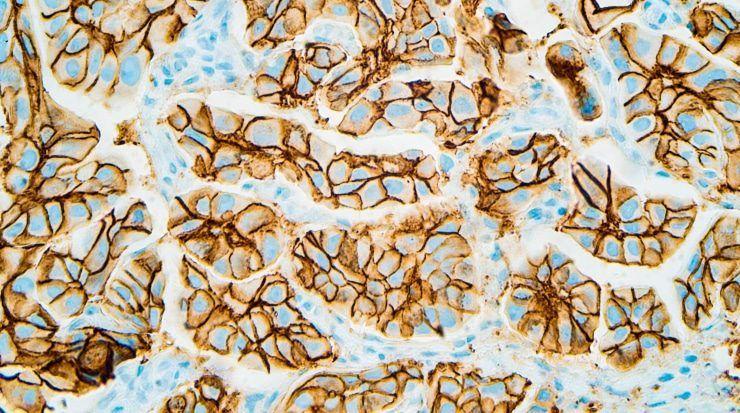
Immunohistochemistry (IHC) is a technique that uses enzyme- or fluorophore-labeled antibodies for visualizing specific antigens in tissue sections. Unlike other types of immunoassays, it preserves the structure and composition of sample material to provide insights into antigen distribution relative to other targets of interest. For this reason, IHC is especially helpful for studying abnormalities such as those observed during cancer or autoimmune disease.

Immunohistochemistry Overview
Immunohistochemistry was first reported in 1942, when Coons et al. used a fluorescein isothiocyanate (FITC) labeled antibody to visualize Pneumococcal bacteria in infected tissue samples. Since then, IHC has benefited from improvements to tissue fixation and sectioning methods, epitope retrieval, and antibody labeling technologies, as well as from the development of advanced microscopy platforms. As a result, IHC is now widely used for applications spanning basic research through to clinical testing.
Uses of Immunohistochemistry
In a research setting, IHC is used for studying proteins of interest under conditions of both health and disease. For example, IHC allows researchers to determine whether protein expression is up-regulated in breast cancer compared to normal tissue, investigate protein mislocalization in cardiovascular disease, and confirm which tissues are targeted during viral infection. In addition, IHC is used for investigating normal physiological processes such as tissue and organ development, wound healing, and programmed cell death to determine how these unfold.
Clinical applications of IHC include its use to identify pathogenic features such as neoplasia, metastasis or inflammation, which can be used for diagnosing and staging disease. IHC is also used for analyzing post-mortem samples, such as the brain tissue of Alzheimer's disease patients, to better understand key pathological mechanisms.
Immunohistochemistry Steps
A typical IHC workflow involves a series of steps, beginning with sample collection. The samples are then embedded to allow for sectioning, before being subjected to (optional) epitope retrieval. Following this, immunostaining is performed using either direct or indirect detection. The tissue sections are then mounted to allow for microscopy-based visualization and analysis.
-
Immunohistochemistry Sample Preparation
IHC sample preparation consists of tissue preservation, followed by embedding and sectioning, and optional epitope retrieval. Only once these steps have been performed can the samples be immunostained for proteins of interest.
- Tissue preservation
Tissue samples destined for immunohistochemical analysis are preserved in one of two ways. Where there is a need to conserve enzyme function or safeguard the antigenicity of sensitive epitopes (e.g., phosphorylated proteins or post-translational modifications), tissue is typically snap frozen by immersion in liquid nitrogen. Where it is critical for tissue morphology to be maintained, formalin fixation followed by paraffin-embedding (often referred to as FFPE) is more commonly used. Although preparing FFPE tissue samples takes longer than cryopreservation (involving up to 24 hours fixation, followed by tissue dehydration and embedding in molten paraffin), it provides the advantage of enabling long-term storage at room temperature.
-
Tissue Embedding and Sectioning
Following preservation, the tissue samples must be sectioned. In the case of frozen material, this is achieved by embedding each sample in Optimal Cutting Temperature (OCT) compound (a water-soluble mixture of glycols and resins) to stabilize it before cutting it into thin slices using a cryostat. The sections are then applied to glass slides prior to being fixed, usually with alcohol or acetone.
FFPE tissue sections, which are already embedded in paraffin, are instead prepared using a microtome. Once applied to glass slides, FFPE sections require deparaffinization and rehydration, and may additionally necessitate epitope retrieval.
Our Gelatin Subbed Slides safeguard tissue attachment throughout the entire IHC workflow and are suitable for use with both frozen and FFPE tissue sections.
-
Epitope Retrieval
Where tissue is fixed using formalin, the formation of protein cross-links can mask epitopes to prevent antibody binding. Formalin-fixed tissue samples may therefore require that an epitope retrieval step be performed. The two main epitope retrieval methods are heat-induced epitope retrieval (HIER), which uses heat and an optimized buffer (typically a citrate buffer of pH 6) for unmasking; and proteolytic-induced epitope retrieval (PIER), which relies on proteases such as proteinase K, trypsin, and pepsin. The chosen epitope retrieval method will be largely dictated by the target and should be carefully optimized during assay development. In many cases, HIER is a preferred choice since it is gentler and more easily controlled.
-
Immunostaining
-
Permeabilization and blocking for IHC
If the antigen of interest is intracellular (e.g., a cytoskeletal protein or a protein associated with a specific subcellular organelle), samples must be permeabilized for antibody reagents to access their target. Permeabilization involves incubating the tissue sections in a mild detergent such as Tween® 20, or a more stringent detergent like Triton™ X-100, or may also be achieved using a solvent such as methanol.
Where the detection method will use horseradish peroxidase (HRP), any endogenous peroxidase activity should be quenched using a dilute solution of hydrogen peroxide. Non-specific binding sites should then be blocked using an appropriate animal serum; it is advised that the serum be matched to the host species of the secondary antibody (e.g., when using a goat anti-mouse secondary, goat serum is recommended for blocking).
We offer a broad range of animal sera for blocking, including sera from donkey, rabbit and goat.
-
Considerations for IHC antibody staining
As for any immunoassay, antibody considerations for IHC include whether to perform direct or indirect immunostaining and the choice of assay readout. Direct staining uses labeled primary antibodies, meaning shorter workflows, while indirect staining uses labeled secondary antibodies and offers signal amplification. For indirect staining, secondary antibodies should be cross-adsorbed against the host species of the sample material to avoid generating unwanted background signal.
Our secondary antibodies are cross-adsorbed against a broad range of serum proteins and immunoglobulins and our donkey secondaries are especially popular for IHC.
Chromogenic detection relies on enzymes such as HRP or alkaline phosphatase (AP) to produce a colored precipitate from specialized IHC substrates and is an established and widely-used approach. Fluorescence-based detection introduces the option for multiplexing. Whichever antibody reagents are selected, it is essential that they are validated for the IHC application and that their use is rigorously optimized in-house using suitable controls.
For help with choosing the right secondary antibody for your research, check out our Secondary Antibody Selection Guide.
-
Mounting and Visualization
Once immunostaining is complete, slides require mounting before being visualized using a microscope. As well as enhancing the quality of the resultant image, mounting helps preserve the sample for viewing at a later date. A broad range of mounting media is available, with many products including a counter-stain such as 4′,6-diamidino-2-phenylindole (DAPI) or propidium iodide (PI) that enable visualization of nuclei.
Our mounting media products include Fluoromount-G®, which boasts over 20,000 literature citations, and DAPI Fluoromount-G®, which includes DAPI for nuclear analysis. We also offer Fluoromount-G® Anti-Fade for applications where preserving fluorescence intensity is critical.
SouthernBiotech is committed to producing and providing the highest quality reagents and we currently offer more than 1,500 antibodies that have been validated for IHC. Contact us today to learn how we can support your research.


